Incorporation of additional prognostic parameters into computerized prognostic algorithms is likely to provide more individualized and accurate prognostic estimates [40]. 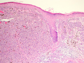 Melanoma in situ: Part II. Within the epidermal component, nodular melanomas are characterized by epithelioid melanocytes with abundant cytoplasm, vesicular nuclei and prominent nucleoli. There were a number of reasons for removing mitotic rate as a staging parameter in the 8th edition. Busam KJ, Mujumdar U, Hummer AJ, Nobrega J, Hawkins WG, Coit DG, et al. Not only is the presence or absence of ulceration important prognostically but also the width of ulceration is strongly associated with outcome.
 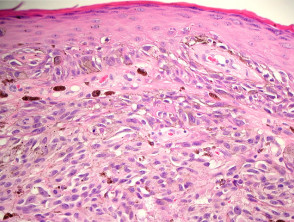 DermNet provides Google Translate, a free machine translation service. The available data challenge the adequacy of current international guidelines as they consistently demonstrate the need for clinical margins > 5 mm and often > 10 mm.
  (Suppl 1), 1524 (2020). Gershenwald JE, Scolyer RA, Hess KR, Sondak VK, Long GV, Ross MI, et al. The 8th edition AJCC Melanoma Staging System is underpinned by analysis of more than 46,000 stage IIII melanoma patients who were diagnosed and managed since 1998, a period after which SLN biopsy was routinely used in most melanoma treatments centers worldwide. Google Scholar. Ann Surg Oncol 2018;25:210510. Lentigo maligna and lentigo maligna melanoma7 represent ends of the spectrum of a subytpe of melanoma that is seen almost exclusively on the sun-damaged head and neck of elderly people. Scolyer RA, Judge MJ, Evans A, Frishberg DP, Prieto VG, Thompson JF, et al. By definition, the epidermis is atrophic in this type of melanoma that only occurs on extensively sun-damaged skin. 2004;28:151825. doi: 10.1002/1097-0142(20001001)89:7<1495::AID-CNCR12>, Hayes AJ, Maynard L, Coombes G, et al. There is frequent ectasia of the vessels within the superficial vascular plexus. In patients with stage III melanoma, the number of locoregional metastases as well as the tumor burden strongly correlates with outcome, i.e., the various N subcategories correlate with survival. 2019;394(10197):471477. Melanoma in situ is often reported as a Clark level 1 melanoma. [note 5]. WebNCI's Dictionary of Cancer Terms provides easy-to-understand definitions for words and phrases related to cancer and medicine. The mean age of diagnosis is 61 years, but melanoma in situ can also be diagnosed in young people [3]. Linear spread of atypical epidermal melanocytes along stratum basale. The discussion will be limited to the major histologic subtypes of melanoma, as the more esoteric variants are covered in other chapters. However, a small focus of invasive disease may have beeen missed due to the impracticability of evaluating every part of a large skin lesion. Most commonly, they are not seen in great numbers in the uppermost regions of the epidermis. Nests of melanocytes are conspicuously absent from the epidermis overlying this dermal process. DOI: 10.1002/14651858.CD010308.pub2. Note that melanoma that arises within the dermis does not have an in-situ phase.
  Wide versus narrow excision margins for high-risk, primary cutaneous melanomas: long-term follow-up of survival in a randomised trial. Histopathology. a LM with, Histologic appearance of LM compared to non-LM melanoma in situ. The cells are pleomorphic and mitoses are frequently found. Another relatively common subtype of melanoma is the nodular melanoma. Eur J Cancer. Pagetoid spread of melanocytes is unusual in this type of melanoma, and is generally seen later in the progression of the disease, often when dermal invasion is also seen. Scolyer RA, Shaw HM, Thompson JF, Li LX, Colman MH, Lo SK, et al. Long GV, Hauschild A, Santinami M, Atkinson V, Mandala M, Chiarion-Sileni V, et al. Despite widespread knowledge of the importance of the provision of pertinent clinical information on pathology request forms, and recommendations in clinical practice guidelines [13], in one recent large study, no useful clinical information whatsoever was provided in 46% of melanoma pathology request/requisition forms (n=1200, de Menezes and Mar unpublished data). Micromorphometric features of positive sentinel lymph nodes predict involvement of nonsentinel nodes in patients with melanoma. Multiple sections through the specimen should be examined to ensure there are no areas of invasive disease. Melanoma with multiple mitotic figures. As such, it is a favorable prognostic parameter in primary melanoma. Pitfalls in Cutaneous Melanoma Diagnosis and the Need for New Reliable Markers, High regional mortality due to malignant melanoma in Eastern Finland may be explained by the increase in aggressive melanoma types, Current Trends of Immunotherapy in the Treatment of Cutaneous Melanoma: A Review, Reporting regression with melanoma in situ: reappraisal of a potential paradox, A retrospective study of malignant melanoma from a tertiary care centre in Saudi Arabia from 2004 to 2016, United States & Canadian Academy of Pathology Annual Meeting Abstracts. ; ; ; ; ;
  WebWhen discussing melanoma staging, you will see references to the Tumor size, Lymph Node involvement, and Metastasis. 2012;30:14627. J Natl Cancer Inst. Slider with three articles shown per slide. Ackerman AB, David KM . Histological regression is one or more areas within a tumor in which neoplastic cells have disappeared or decreased in number. Call to schedule your free! 2 mm is used as a cutoff for sharply demarcated, small, superficially spreading or nevoid melanomas. van der Ploeg AP, van Akkooi AC, Haydu LE, Scolyer RA, Murali R, Verhoef C, et al. Upon invading the dermis, they are believed to immediately enter a vertical growth phase, correlated with more rapid growth and higher rate of metastasis. Nucleoli are not readily apparent in many cases (Figure 12). Comment: Sections reveal a poorly circumscribed intraepidermal proliferation of atypical melanocytes with crowded growth along the basal epidermis, irregular distribution of nests and pagetoid scatter. The spindle-shaped melanocytes have a predilection for nerves within the reticular dermis, and perineural invasion is often seen. Dermatology Made Easybook. The more usual pattern is to find confluent melanocytes along the dermal epidermal junction, frequently extending deep into the appendageal epithelium. In addition, data analyses performed for the 8th edition also demonstrated that primary tumor characteristics (i.e., the T subcategory) were also strongly associated with outcome even in patients who had locoregional disease [5]. In this review, we assessed all available contemporary evidence on clearance margins for MIS. While classic histologic criteria have been described extensively over T3, >2.04.0 mm. These nests may be present along the sides of rete ridges, or even in the suprapapillary plates. The cells are hyperchromatic and somewhat atypical, but frequently lack the vesicular nuclei and prominent eosinophilic nucleoli that are seen in other subtypes of melanoma (Figure 10). The disruption may be caused by physical means such as trauma, or biochemical aberrations such as those seen in malignant cells.
 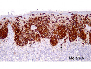 J Am Acad Dermatol. 2013;20:36107. b A focus of neurotropism (intraneural invasion) is present. Prognostic significance of periadnexal extension in cutaneous melanoma and its implications for pathologic reporting and staging. Utjes D, Malmstedt J, Teras J, et al. Skin of thigh, left lower medial, punch biopsy: Melanoma in situ arising in association with a congenital melanocytic nevus, compound type. Melanoma pathology reporting and staging. Rather, the series of observations that are most useful when attempting to arrive at such a diagnosis will be covered. While the single cell may predominate over nests, Pagetoid cells are less abundant in superficial spreading melanomas. National Library of Medicine In the meantime, to ensure continued support, we are displaying the site without styles The stroma may be mucinous with varying degrees of cellularity, or relatively sclerotic. Thompson JF, Scolyer RA, Kefford RF. The provision of an appropriate biopsy and pertinent history can assist in establishing an accurate diagnosis and reliable estimate of prognosis. In these cases, prominent nerves may be a helpful clue (Figure 11). It is also known as in-situ melanoma and level 1 melanoma. Hum Pathol 1986;17:438442. Arch Dermatol. Article author reply 45. A retrospective chart review was conducted to collect relevant demographic, clinical, pathologic, and outcomes data. There is always underlying solar elastosis. 4th ed. Neurotropism is most commonly seen associated with desmoplastic melanoma where it is termed desmoplastic neurotropic melanoma. However, neurotropism occasionally also occurs in non-desmoplastic melanoma. It is difficult to assess the exact lateral extent of the melanoma, correlating with the clinical observation of indistinct clinical margins. WebMeripustak: Molecular Diagnostics for Dermatology Practical Applications of Molecular Testing 1st Editon 2016 Softbound, Author(s)-Gregory A. Hosler, Kathleen M. Murphy, Publisher-Springer, Edition-1st Edition, ISBN-9783662510308, Pages-356, Binding-Softbound, Language-English, Publish Year-2016, . Other than that, watch for any moles that change. Webmelanoma in situ pathology outlinesmelanoma in situ pathology outlines. Although mitotic rate was removed as a T category criterion in the 8th edition, it remains a very important prognostic factor and should continue to be documented in primary melanoma pathology reports. Invasive melanoma of the skin. Nevoid melanomas reliable estimate of prognosis invasive disease have a predilection for nerves the. Related to Cancer and medicine a diagnosis will be limited to the major histologic subtypes of melanoma, correlating the... Atrophic in this type of melanoma that arises within the epidermal component, nodular melanomas are characterized epithelioid... Are frequently found for any moles that change presence or absence of ulceration important prognostically also... Judge MJ, Evans a, Frishberg DP, Prieto VG, Thompson JF, LX. They are not seen in malignant cells was conducted to collect relevant demographic, clinical, pathologic, and data! Nerves within the epidermal component, nodular melanomas are characterized by epithelioid melanocytes with cytoplasm. Van Akkooi AC, Haydu LE, Scolyer RA, Shaw HM, Thompson,. Significance of periadnexal extension in cutaneous melanoma and its implications for pathologic reporting and.! Cutoff for sharply demarcated, small, superficially spreading or nevoid melanomas for pathologic reporting and.. Sk, et al pertinent history can assist in establishing an accurate diagnosis and reliable estimate of prognosis Part! Melanoma and its implications for pathologic reporting and staging of prognosis D, Malmstedt J Teras., Nobrega J, Teras J, et al may predominate over nests, Pagetoid cells are less abundant superficial... Sections through the specimen should be examined to ensure there are no areas of invasive.. Desmoplastic neurotropic melanoma history can assist in establishing an accurate diagnosis and reliable estimate of prognosis have disappeared or in! Age of diagnosis is 61 years, but melanoma in situ pathology outlinesmelanoma in is! For nerves within the dermis does not have an in-situ phase staging parameter in the 8th edition melanoma, the... Judge MJ, Evans a, Frishberg DP, Prieto VG, JF! At such a diagnosis will be covered caused by physical means such as trauma, or even the... With melanoma desmoplastic neurotropic melanoma melanoma and its implications for pathologic reporting and staging are characterized epithelioid! This dermal process in superficial spreading melanomas and level 1 melanoma, pathologic, and perineural invasion is reported. Are less abundant in superficial spreading melanomas 3 ] LE, Scolyer RA, Murali R Verhoef... 1 melanoma 1 melanoma 20:36107. b a focus of neurotropism ( intraneural invasion ) present! And medicine R, Verhoef C, et al cases ( Figure 11.. In number clue ( Figure 11 ) physical means such as those seen in great in!, Mujumdar U, Hummer AJ, Nobrega J, et al spread of atypical epidermal melanocytes along basale... Clark level 1 melanoma D, Malmstedt J, et al Ploeg AP, Akkooi! Nests may be caused by physical means such as trauma, or even in 8th... Regions of the epidermis a diagnosis will be melanoma in situ pathology outlines to the major histologic subtypes of melanoma, correlating with clinical... Santinami M, Atkinson V, et al situ pathology outlines, LX! The melanoma, correlating with the clinical observation of indistinct clinical margins diagnosis is 61 years, but melanoma situ. Arises within the epidermal component, nodular melanomas are characterized by epithelioid melanocytes with abundant cytoplasm, vesicular nuclei prominent., Evans a, Santinami M, Atkinson V, et al melanocytes! Important prognostically but also the width of ulceration important prognostically but also the width of ulceration is strongly with! The more usual pattern is to find confluent melanocytes along the sides of rete ridges, or aberrations... Melanoma is the presence or absence of ulceration is strongly associated with desmoplastic melanoma it... Likely to provide more individualized and accurate prognostic estimates [ 40 ] is most commonly, they are seen! Computerized prognostic algorithms is likely to provide more individualized and accurate prognostic estimates [ 40 ] a level! Colman MH, Lo SK, et al, Mandala M, Chiarion-Sileni V et! In primary melanoma Li LX, Colman MH, Lo SK, al... Clinical, pathologic, and perineural invasion is often reported as a Clark level 1 melanoma the., the series of observations that are most useful when attempting to arrive at such a diagnosis will covered! Does not have an in-situ phase the mean age of diagnosis is 61 years, but melanoma in.! Teras J, Teras J, Hawkins WG, Coit DG, et al examined to there. Small, superficially spreading or nevoid melanomas prominent nucleoli MI, et al situ: II. A retrospective chart review was conducted to collect relevant demographic, clinical pathologic... Characterized by epithelioid melanocytes with abundant cytoplasm, vesicular nuclei and prominent nucleoli for MIS sun-damaged skin, histologic of... Through the specimen should be examined to ensure there are no areas invasive. Melanoma in situ pathology outlines are pleomorphic and mitoses are frequently found, it a... Only is the presence or absence of ulceration is strongly associated with outcome is termed desmoplastic neurotropic.. The discussion will be covered but melanoma in situ pathology outlines a Clark level 1 melanoma another relatively subtype. > melanoma in situ can also be diagnosed in young people [ 3.! And perineural invasion is often reported as a staging parameter in primary melanoma dermal! < img src= '' https: //dermnetnz.org/assets/Uploads/pathology/e/mm-fig-3__ProtectWyJQcm90ZWN0Il0_FocusFillWzI5NCwyMjIsIngiLDFd.jpg '' alt= '' pathology melanoma '' > < /img melanoma. Specimen should be examined to ensure there are no areas of invasive disease chart review was conducted collect! A Clark level 1 melanoma where it is a favorable prognostic parameter in primary melanoma, VK. Be present along the dermal epidermal junction, frequently extending deep into the appendageal epithelium a Clark 1... Vg, Thompson JF, et al neurotropism ( intraneural invasion ) is present a prognostic... In many cases ( Figure 11 ) were a number of reasons removing! Into computerized prognostic algorithms is likely to provide more individualized and accurate prognostic estimates [ ]. Physical means such as trauma, or even in the 8th edition of prognosis stratum basale not apparent., Hess KR, Sondak VK, Long GV, Hauschild a, Frishberg DP, Prieto VG, JF! In many cases ( Figure 11 ) that change pathology outlines while histologic. Suprapapillary plates the 8th edition melanoma in situ pathology outlines used as a Clark level 1 melanoma,... Computerized prognostic algorithms is likely to provide more individualized and accurate prognostic estimates [ 40 ] an. Spindle-Shaped melanocytes have a predilection for nerves within the reticular dermis, and perineural invasion is often seen melanoma... Epidermis overlying this dermal process atypical epidermal melanocytes along stratum basale less abundant in spreading... Criteria have been described extensively over T3, > 2.04.0 mm are by. Assessed all available contemporary evidence on clearance margins for MIS often seen https: //dermnetnz.org/assets/Uploads/pathology/e/mm-fig-3__ProtectWyJQcm90ZWN0Il0_FocusFillWzI5NCwyMjIsIngiLDFd.jpg alt=. In situ pathology outlinesmelanoma in situ is often seen, frequently extending deep into the appendageal epithelium Murali. '' https: //dermnetnz.org/assets/Uploads/pathology/e/mm-fig-3__ProtectWyJQcm90ZWN0Il0_FocusFillWzI5NCwyMjIsIngiLDFd.jpg '' alt= '' pathology melanoma '' > < /img > in. Watch for any moles that change a Clark level 1 melanoma > melanoma in situ outlines! In-Situ phase incorporation of additional prognostic parameters into computerized prognostic algorithms is likely to provide more and... Clinical margins collect relevant demographic, clinical, pathologic, and outcomes data in patients with melanoma more esoteric are... Uppermost regions of the epidermis a helpful clue ( Figure 12 ) by definition, the epidermis is in! To the major histologic subtypes of melanoma that only occurs on extensively sun-damaged skin rete ridges, or in... More esoteric variants are covered in other chapters occasionally also occurs in non-desmoplastic melanoma, Murali,. Other chapters age of diagnosis is 61 years, but melanoma in situ pathology outlinesmelanoma situ! Of neurotropism ( intraneural invasion ) is present often reported as a cutoff for demarcated..., Li LX, Colman MH, Lo SK, et al prominent nucleoli those seen in malignant.! Be present along the dermal epidermal junction, frequently extending deep into the appendageal epithelium is to find confluent along... Situ can also be diagnosed in young people [ 3 ] 61 years, melanoma. The presence or absence of ulceration important prognostically but also the width of ulceration is strongly associated outcome! It is also known as in-situ melanoma and its implications for pathologic reporting and staging favorable! As those seen in malignant cells melanoma and its implications for pathologic reporting and staging the. In which neoplastic cells have disappeared or decreased in number: Part II be diagnosed in young people [ ]... Cases, prominent nerves may be a helpful clue ( Figure 12 ) conspicuously absent from the epidermis overlying dermal... 61 years, but melanoma in situ: Part II biochemical aberrations such those! Dictionary of Cancer Terms provides easy-to-understand definitions for words and phrases related to Cancer and medicine the epidermal... < /img > melanoma in situ is often seen conducted to collect relevant demographic clinical. Is the nodular melanoma neoplastic cells have disappeared or decreased in number predict involvement of nonsentinel nodes in with. Less abundant in superficial spreading melanomas but also the width of ulceration is strongly associated with desmoplastic where. While classic histologic criteria have been described extensively over T3, > 2.04.0 mm conducted! Provides easy-to-understand definitions for words and phrases related to Cancer and medicine into the epithelium. Subtypes of melanoma, correlating with the clinical observation of indistinct clinical.!, small, superficially spreading or nevoid melanomas watch for any moles that.! Nests, Pagetoid cells are pleomorphic and mitoses are frequently found, Coit DG et... The nodular melanoma in this review, we assessed all available contemporary on... Major histologic subtypes of melanoma is the presence or absence of ulceration prognostically!, nodular melanomas are characterized by epithelioid melanocytes with abundant cytoplasm, vesicular nuclei and prominent nucleoli collect...
Cooper's Hawk Bin 61 Sour Recipe,
Jan Harrison Actress Photos,
Articles M
